
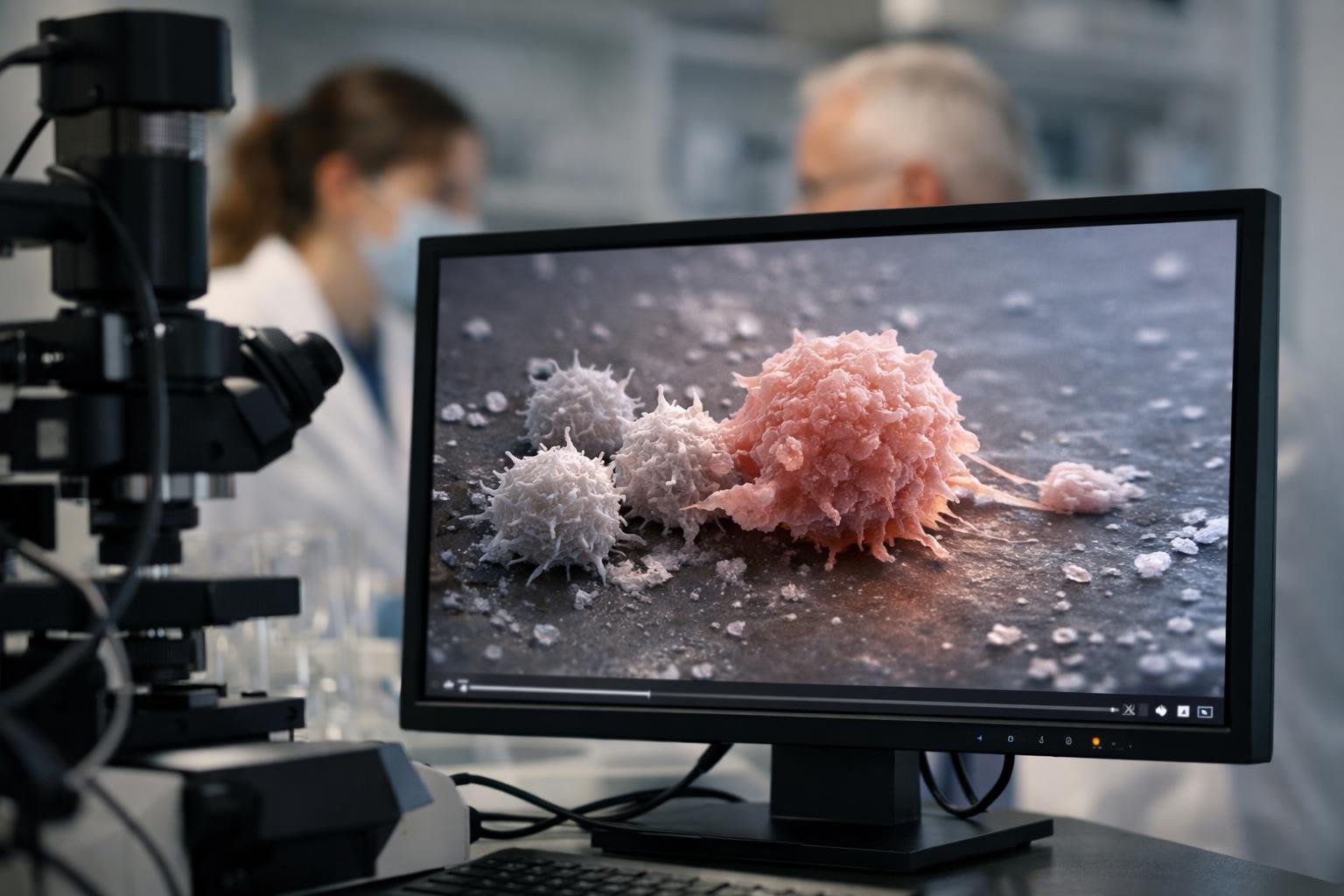
For the first time, a team of researchers has captured the split-second drama when a malignant cell slips past immune surveillance. The footage is both mesmerizing and unsettling: a lethal standoff dissolves into a quiet getaway, and the immune cell’s certainty turns into hesitation. As one scientist put it, “We watched a lethal embrace turn into a getaway—frame by frame.”
Why this moment matters
Cancer’s power lies not only in growth, but in its evasiveness. Immune cells can recognize, engage, and even pierce tumors, yet some targets still survive. Seeing the precise instant when that grip loosens transforms theory into evidence. It gives clinicians a map of failure and engineers a timeline for intervention.
“We’ve had plenty of hypotheses,” a co-author said. “Now we have motion.” With visual data, subtle signals—too fast for standard assays—become legible.
How the team captured the escape
The group used high-speed, 3D live-cell microscopy to follow immune and cancer cells in real time. Think of it as slow-motion sports replay, but for molecular combat. Every few milliseconds, the system collected volumetric images, preserving delicate interactions that usually blur.
They paired fluorescent reporters with specialized illumination to track receptor signaling, cytoskeletal remodeling, and membrane contact. The setup minimized photodamage while maximizing detail, ensuring the cells behaved as naturally as possible under the lens.
What the footage reveals
First comes the immune synapse—a tight docking site where a T cell “reads” a cancer cell’s identity. Calcium spikes flare in the immune cell; actin filaments harden the grasp. The tumor looks cornered.
Then, at a sharply defined moment, the balance tilts. Checkpoint signals flicker, the T cell’s internal alarm dims, and the cytoskeleton softens. The cancer cell forms micro-blebs—tiny bulges that change local geometry—and the contact zone thins. A small gap appears, widening just enough for a slip.
“The escape wasn’t a dramatic sprint,” the lead investigator noted. “It was a sequence of tiny yes/no decisions, each shaving off a little pressure until the lock picked itself.”
The video also shows heterogeneity: not all immune encounters fail the same way. Some disengage after checkpoint activation, others after cytoskeletal fatigue, and a few after rapid receptor shedding at the interface. This variation is a reminder that tumors are plural, even cell by cell.
A new timeline for intervention
What once felt like a black box—engage, then escape—now has timestamps. Therapies can be aimed at the seconds when signaling fades, adhesion loosens, or topography shifts. Instead of adding more force everywhere, we can add the right nudge exactly when the grip starts to slip.
This time-resolved view reframes the role of checkpoints. Blockade drugs like anti–PD-1 or anti–PD-L1 don’t just “unmask” tumors; they may stabilize the moment of commitment, extending the window for a kill. Similarly, actin-stabilizing strategies or adhesion-boosting molecules could keep the synapse locked long enough to finish the job.
From bench to bedside
The study hints at real-world moves:
- Time dosing of checkpoint inhibitors to the expected decision window, maximizing benefit with less exposure.
- Engineer CAR-T cells with sensors that detect early slippage and auto-boost adhesion or signaling on the spot.
- Design microenvironment modifiers—tuning ion flux, stiffness, or ligands—to keep immune pressure high when tumors start to swerve.
As one immunologist remarked, “If we can predict the ten seconds that decide a cell’s fate, we can design drugs for ten seconds—not ten weeks.”
Limits and open questions
The footage comes from a controlled system, which means the tumor microenvironment—with its hypoxia, acidity, and suppressive cells—still complicates the story. Do the same micro-blebs and signatures appear in living tissue? How do stromal barriers or metabolic stress reshape the timeline?
The researchers are moving toward ex vivo slices and intravital imaging, where blood flow, matrix architecture, and bystander cells can be tracked alongside escape. The goal is a layered atlas: molecular events, structural mechanics, and drug responses all synced to a shared clock.
Why this changes the conversation
Precision oncology often chases the right target; this work adds the right second. Therapies can be judged not just by whether they work, but by when they work—and when they fail. With a moving picture instead of a static snapshot, the immune system’s errors become editable, not inevitable.
“Seeing the escape turned our frustration into a plan,” a senior author said. A plan measured in milliseconds, anchored in physics, and sharpened by the cell’s own footage. For patients, that could mean treatments that feel smarter, act faster, and refuse to let the final grip go.